Shoulder pain is one of the most common complaints after motor vehicle collisions, sports injuries, or repetitive strain. Many patients assume they need an MRI right away, but in reality, an accurate diagnosis often begins with choosing the correct X-ray view. One of the most valuable but frequently overlooked imaging tools is the transcapular Y-view, also called the lateral scapular Y-view.
This specialized view can reveal important structural details that a standard shoulder X-ray simply cannot show. In our clinic, we regularly use this imaging technique to identify conditions such as shoulder impingement, rotator cuff involvement, or abnormal positioning of the humeral head.
Below is a real example that highlights why this view matters and how clinical competency in imaging directly impacts a patient’s diagnosis and care.
What Makes the Y-View Different
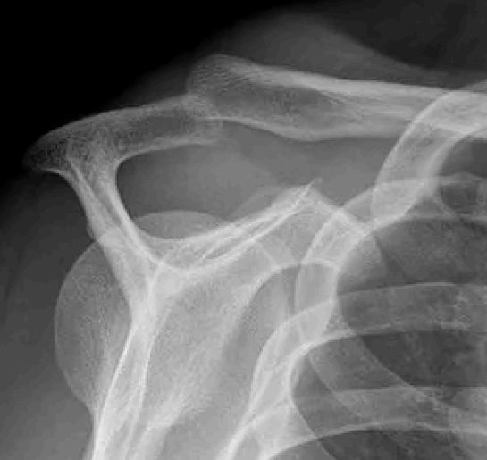
Most people are familiar with the standard front-to-back shoulder X-ray. It shows bones clearly but does not always reveal what is happening in the subacromial space, where tendons like the supraspinatus pass. The Y-view, however, is taken at an angle through the scapula so the acromion, coracoid, and scapular body form a “Y” shape on the image.
This view provides a clear visualization of:
The alignment of the humeral head in relation to the glenoid
The contour of the acromion
The subacromial and subclavicular spaces
The path of the supraspinatus tendon as it inserts into the humerus
Because of this positioning, the Y-view offers critical information when evaluating patients with suspected impingement or mechanical shoulder pain. It helps clinicians pinpoint whether structural narrowing or altered biomechanics are contributing to the patient’s symptoms.
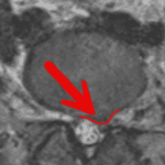
On pages 2–3 of the original case report, the X-ray images clearly show how the supraspinatus tendon passes beneath the acromion and clavicle, providing a much more meaningful view of the space involved in impingement cases.
A Case Example: Getting the Right Picture
A patient presented with classic signs of shoulder impingement. Their clinical exam included painful arc testing, positive orthopedic tests, and limited range of motion. Rather than sending the patient immediately for advanced imaging, a transcapular Y-view was obtained.
This single X-ray view provided essential diagnostic information:
It visualized the subacromial region with clarity.
It illustrated how the humeral head was interacting with the acromion.
It supported the clinical findings pointing toward supraspinatus involvement.
By combining the physical examination with the Y-view, we were able to confirm the diagnosis and determine appropriate next steps. This is the type of clinical reasoning and imaging selection that prevents misdiagnosis and ensures the treatment plan matches the actual pathology.
Why This Matters for Patients and Attorneys
For patients, getting the right diagnosis early means fewer delays, fewer unnecessary procedures, and a clearer path to recovery. For attorneys handling motor vehicle or personal injury cases, accurate imaging and documentation are essential in demonstrating the nature and severity of injuries.
The Y-view is not a “replacement” for MRI, nor is it intended to rule out soft tissue injury. Instead, it is a precision tool that adds valuable, cost-effective information and helps guide additional imaging when appropriate.
Unfortunately, the Y-view is often overlooked in general practice. Many clinics rely solely on standard views, which can miss subtle but important details. This is where training, experience, and advanced imaging competency make a difference.
Our Approach at Eatontown Elite Care Center
As a Fellowship-trained Primary Spine Care provider, I place significant emphasis on choosing the correct imaging for each case. The Y-view is a prime example of why proper imaging technique matters. It provides insight into mechanical causes of shoulder pain that simply do not appear on standard X-rays.
When patients come to our office after an accident or injury, they can expect:
A careful, evidence-based examination
Expertise in selecting and interpreting the right imaging views
Clear communication with their legal and medical teams
Accurate, defensible documentation for both treatment and case support
Advanced imaging knowledge is one of the most meaningful ways we improve outcomes and ensure every patient receives the highest level of care.